Workflows
Structural Retinal Degeneration
In rd10 or other RP lines imaged with the Envisu, you will see a progressive thinning of the outer retina while the inner retinal layers (like the ganglion cell layer and inner plexiform layer) remain relatively preserved in the early-to-mid stages of the disease.
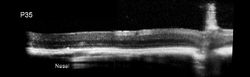
SD-OCT imaging of wild-type mice demonstrated visibility of the inner segment/outer segment (IS/OS) junction, external limiting membrane (ELM), outer nuclear layer (ONL), and outer plexiform layer (OPL). At P14, most rd10 mice exhibited normal SD-OCT profiles, but some displayed changes in the IS/OS junction. At the same time point, rd1 mice had severe outer retinal degeneration. In rd10 mice, imaging revealed loss of the IS/OS junction by P18, hyperreflective changes in the ONL at P20, hyperreflective vitreous opacities, and shallow separation of the neural retina from the RPE.
 |  |  |  |  |  |
|---|---|---|---|---|---|
 |
Retinal Vascular Changes
I'm a paragraph. Click here to add your own text and edit me. It's easy.
| | | | | |
|---|---|---|---|---|---|
|